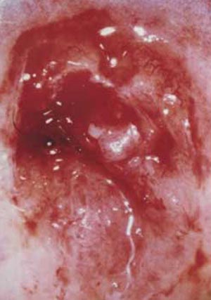
Uterine Cervix Cancer

Cancer of the uterine cervix is a condition when the cells of the cervix undergo abnormal growth. Early detection is the key to treatment and cure. Because of the slow progression of cancer of the cervix, it can be detected early and thereby prevented. We examine the role of pap smears in examining the cervical cells for abnormalities that can be indicative of malignancy. Cervix biopsies of different types are diagnostic procedures that can also function as treatments for pre-cancerous lesion in the cervix area.
Cancer uterine cervix
The uterine cervix is located at the lower part of the uterus and forms a canal that opens into the vagina. Uterine cervix cancer does not cause any pain or other symptoms in its early stages. It is only when the abnormal cervical cells become cancerous that abnormal bleeding occurs. Increased vaginal discharge may also be another symptom. Other symptoms of cervix cancer are ulcers in the cervix and bleeding after intercourse. Cervix cancer does not spread early. It spreads by way of the lymphatic system. Cervical cancer tends to be slow growing.
There is a tendency for cells to appear on the surface of the cervix. These cells are not always cancerous. But they must be observed for any abnormality since it might be indicative of cancer. Cancer of the cervix can be best detected by the pap smear that detects changes on the cervix before it becomes malignant. A pap smear helps in detecting any abnormality in the cells of the cervix. Abnormality in the cells of the cervix can be of two types:
Low grade SIL (squamous intraepithelial lesion): This refers to early changes in the size, number and shape of the cells on the surface of the cervix. This condition generally occurs in women in the age group 25 - 45. Though some low grade lesions go away without treatment, some of them may grow larger and form high-grade lesions. Low-grade lesions are treated with cauterization (burning) or laser surgery to destroy the abnormal cells without damaging the nearby healthy tissue. Cryosurgery (freezing) is also sometimes resorted to.
High grade SIL: This condition is characterized by a large number of pre-cancerous cells that look different from normal cells. This is typically seen in women in the age group 30 - 40. Such high grade SIL cells may take years to invade deeper layers of the cervix to become cancerous.
Pap Smear
A pap (Papanicolaou) smear is recommended annually for women over the age of 21, especially for those who have been sexually active. A pap smear is a painless test that is used to detect abnormal cells in and around the cervix. A doctor uses a speculum to widen the vagina and scrapes a few cells from the cervix and upper vagina. A spatula helps in collecting these cells that are then placed on a glass slide and checked for abnormality in a laboratory. This test is ideally conducted between 10 and 20 days after the first day of the menstrual period in a doctor's office or a health clinic. The Bethesda System is used for describing low-grade and high-grads SIL conditions on a pap smear. Class 1 indicates a condition where the cells in the pap smear sample are normal whereas Class 5 is indicative of invasive cancer. A pap test as a screening test shows a positive result for malignant and pre-malignant changes in the cervix. It is indicative of a need for further diagnostic procedures. A pap smear is not a diagnostic tool - it is conducted on women who have so symptoms of cancer. A cervix biopsy is a diagnostic tool.
Cervix biopsy
Colposcopy is used to check the cervix for abnormalities. It involves examination of the cervix under magnification to locate any abnormalities. The Schiller Test is yet another diagnostic tool wherein Iodine solution may be used to coat the cervix. Healthy cervix cells turn brown whereas the abnormal cells turn white or yellow. A cervix biopsy involves removal of small amount of cervical tissue for examination. One procedure involves using an instrument to pinch off small pieces of cervical tissue. The LEEP (Loop Electro surgical Excision Procedure) is yet another technique of cervix biopsy wherein an electric wire loop is used to slice off a thin round piece of tissue. A cone biopsy of the cervix or conization is another procedure that allows pathologists to see the invasion of the abnormal cells in the cervix. This procedure is also used as a treatment for pre-cancerous lesions in the area. When this form of cancer advances, it grows laterally towards the pelvic wall and results in renal failure as it obstructs the ureters. In cases of invasive cervical cancer, chemotherapy, radiation or surgery and hysterectomy are carried out.
The treatment for uterine cervix cancer depends on the location and size of the tumor and its stage of advancement. Staging is an attempt to find out the extent of spread of the disease. Blood and urine tests as well as other procedures such as cystoscopy and proctosigmoidoscopy are used to find out the extent of spread of the disease. Cystoscopy is a procedure whereby the doctor uses a thin, lighted instrument to look inside the bladder. Proctosigmoidoscopy is used to check the rectum and lower parts of the large intestine. Ultrasonography and MRI scans are also frequently used.
Instances of cervical cancer are linked to the infection caused by human papillomavirus (HPV) that is spread by sexual contact. Instances of cervix cancer are seen more in women who have many sexual partners or those with poor genital hygiene or those whose spouses have many sexual partners. Cigarette smoking and oral contraceptives have also been known to increase the risk of developing abnormal changes in the cells of the cervix. The importance of regular pelvic examinations and annual pap smears cannot be ignored.
Top of the Page: Uterine Cervix Cancer
Tags:#cervix #cervix cancer #cervix biopsy #cancer cervix uterine #cervical cancer

Enlarged Uterus
Bacterial Vaginosis
Yeast Infection
Irregular Menstrual Cycle
PMS - Menstruation
Dysmenorrhea
Hypomenorrhea
Mid Cycle Bleeding
Pelvic Organ Prolapse
Vaginal Atrophy
Cervix Cancer
Abnormal Pap Smear
Polycystic Ovarian Syndrome
Ovulation Pain
Uterine Prolapse
Fibroid Tumor
Menorrhagia
Endometriosis Symptom
Galactorrhea
Hysterectomy
Blocked Fallopian Tubes
Menopause and Weight Gain
Premature Menopause
Surgical Menopause
Other health topics in TargetWoman Women Health section:
General Women Health

Women Health Tips - Women Health - key to understanding your health ...
Cardiac Care
Women's Heart Attack Symptoms - Identify heart problems...
Skin Diseases
Stress Hives - Red itchy spots ...
Women Disorders
Endocrine Disorder - Play a key role in overall wellbeing ...
Women's Reproductive Health
Testosterone Cream for Women - Hormone replacement option ...
Pregnancy
Pregnancy - Regulate your lifestyle to accommodate the needs of pregnancy ...
Head and Face
Sinus Infection - Nearly 1 of every 7 Americans suffer from ....
Women and Bone Care

Slipped Disc - Prevent injury, reduce pain ...
Menstrual Disorders
Enlarged Uterus - Uterus larger than normal size ...
Female Urinary Problems
Bladder Problems in Women - Treatable and curable ...
Gastrointestinal Disorders
Causes of Stomach Ulcers - Burning feeling in the gut ...
Respiratory Disorders
Lung function Test - How well do you breathe ...
Sleep Management

Insomnia and Weight Gain - Sleep it off ...
Psychological Disorders in Women
Mood swings and women - Not going crazy ...
Supplements for Women
Women's Vitamins - Wellness needs...
Natural Remedies

Natural Diuretic - Flush out toxins ...
Alternative Therapy
Acupuncture Point - Feel the pins and needles ...
Top of the Page: Uterine Cervix Cancer
Popularity Index: 104,866

